
Fig. 8.
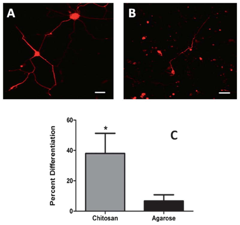
Cortical neuron differentiation within hydrogels. (A) Cortical neuron showing extensive neurite outgrowth within chitosan hydrogels (B) neurite extension within agarose hydrogels indicating low percentage differentiation. Scale bar = 50 μm. (C) Quantification of neurite outgrowth within chitosan (0.5%) and agarose hydrogels (1% Seaprep®). * indicates statistically significant higher percentage (p < 0.05) of neurite extension from chitosan hydrogels as compared to agarose hydrogels.