
Fig. 3. Promotion of invasion and metastasis by NRP2b.
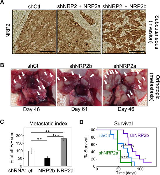
(A) Immunohistochemical staining for NRP2 in tumors derived from control H358 cells (shCtl, left) or shNRP2 cells with reexpression of either NRP2a (middle) or NRP2b (right), grown as subcutaneous xenografts in immunocompromised mice (n = 8 for each condition). (B) Orthotopic model of H358 cells knocked down with shCtl, shNRP2b, or shNRP2a (n = 10 for each condition). Macroscopic deposits of metastatic tumor cells attached to the thoracic cavity (white arrows) were photographed and counted. (C) Metastatic index. Numbers of chest wall metastases/animal formed from shNRP2b and shNRP2a cells were normalized to the time after injection and compared to shControls. Significance was determined by linear combinations of ANOVA parameters analyzed on a log scale. (D) Kaplan-Meier survival analysis of the orthotopic cohorts from (B). Significance was assessed using the log-rank test. In (C) and (D), *P ≤ 0.05; **P ≤ 0.01; ***P ≤ 0.001.