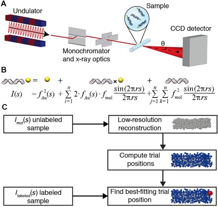
Fig. 1. Schematic of SAXS measurements and workflow to determine gold label positions in macromolecular reconstructions.

(A) Schematic of SAXS measurements. The incident synchrotron-generated x-ray beam (red line) is shown with x-ray optics. Single gold-labeled macromolecules are placed into the x-ray beam in a sample cell. The direct beam is blocked by a beam stop, and scattered photons are detected using a charge-coupled device (CCD) detector. (B) Scattering intensity equation for a single labeled molecule. The scattering signal can be decomposed into a sum of the individual scattering contributions: the gold label scattering, the gold-macromolecule cross-term, and the scattering from the macromolecule only. (C) Schematic of the workflow to determine the positions of gold labels relative to low-resolution three-dimensional (3D) reconstructions of unlabeled macromolecules. The SAXS profile of the unlabeled sample is used in ab initio reconstruction of a low-resolution model consisting of dummy residues or beads. Sterically allowed gold label trial positions are computed in the bead model. Experimental data from the gold-labeled sample are compared to the computed scattering profile for each of the trial positions, and the best-fitting positions are identified.
