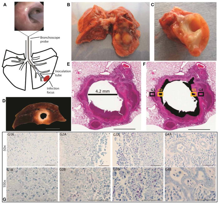
Fig. 1. Disease lesions in the rabbit model of pulmonary cavitary TB.
(A) Diagram showing the method of bronchoscopic Mtb infection of the rabbit lung. Thick black lines show outline of lung lobes, thin black lines show the outline of bronchi, the gray line shows the bronchoscope probe, the blue line shows the inoculation tube, and the red oval shows the infection focus. Top image shows placement of the bronchoscope past the tertiary bronchial division. (B) Freshly resected rabbit lung showing gross pathology at the focus of Mtb infection. (C) Transverse section through the tubercolosis (TB) lesion in the rabbit lung in (B) confirming presence of a cavity. (D) Formalin-fixed cavity in the lower lung lobe of the rabbit model similar to the cavity in (C). (E) Hematoxylin and eosin (H&E) staining of a formalin-fixed representative cavity formed in rabbit lung after bronchoscopic infection with Mtb. The diameter of the cavity after fixation was 4.2 mm. (F) A modification of the same image in (E). The black sections identify areas of necrosis that were grossly identified as caseum (liquefied cell debris) in (D). (G) A fixed tissue section serial to the one in (E). The tissue section was formalin fixed and acid-fast stained; Mtb bacteria are stained red, other cells are blue. (G1A) to (G4A) show four fields of view taken at 50× corresponding to the box insets G1 to G4 in (F). (G1A) and (G4A) show regions of the cavity wall in (F) box insets G1 and G4 (black); (G2A) and (G3A) show regions of caseum/necrosis in (F) box insets G2 and G3 (yellow). (G1B) to (G4B) show four fields of view taken at 100× corresponding to the box insets G1 to G4 in (F). Scale bars, 10 mm (D), 3 mm (E and F), and 50 μm (G).

