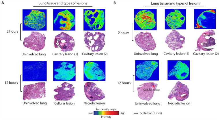
Fig. 4. MALDI-MSI analysis and gross pathology of rabbit lung after multiple doses of rifampin or rifapentine.
Ion density maps of rifampin (A) or rifapentine (B) after multiple dosing in rabbit lung biopsies taken 2 or 12 hours after the final dose are shown. At 12 hours after the final dose, the rifampin signal was higher in necrotic lesions than in surrounding infected lung tissue, whereas rifapentine did not concentrate in the necrotic lesions/caseum. All MALDI-MS images are shown on a fixed ion density scale. Images of H&E-stained lung tissue sections from the same biopsy are shown below each ion density map to confirm gross pathology and the type of lung lesion. Scale bar, 5 mm.

