
Figure 1. G3BP1-APEX2 Mediates Specific Biotinylation of Stress-Granule-Associated Proteins.
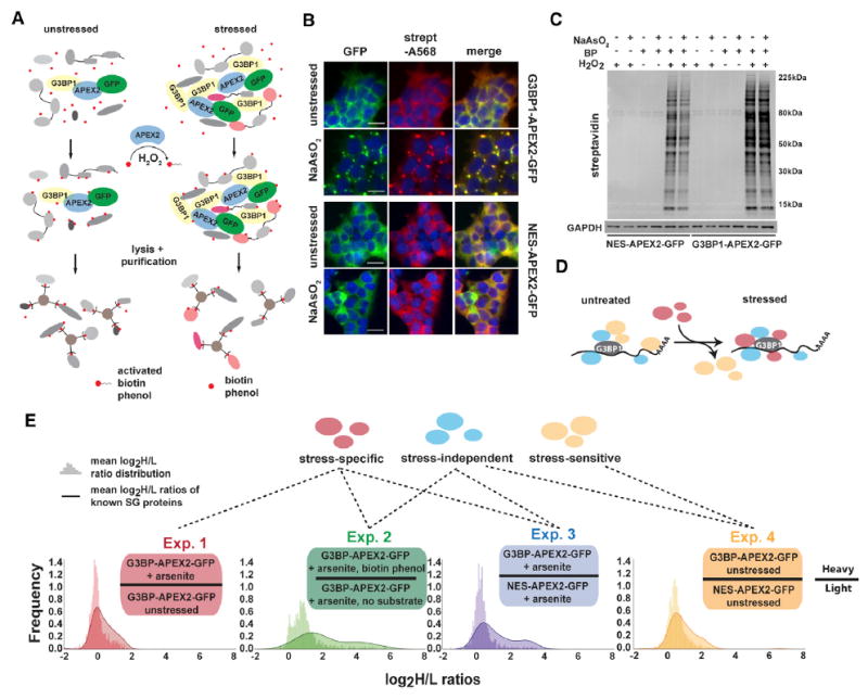
(A) Schematic of APEX proximity labeling to tag SG proteins with biotin.
(B) Streptavidin staining of unstressed and NaAsO2-treated HEK293T G3BP1-APEX2-GFP and hPGK-NES-APEX2-GFP cells. Scale bars, 25 μm.
(C) Streptavidin-HRP western blot analysis of induced protein biotinylation in lysates from NES-APEX2-GFP and G3BP1-APEX2-GFP cells.
(D) Schematic of G3BP1 interactome changes upon stress.
(E) Experimental designs for detecting the G3BP1 interactome changes under different conditions, including log2 H/L ratio distributions of all proteins detected, overlaid with log2 H/L ratio distributions of known SG proteins.
See also Figures S1 and S2 and Table S1.