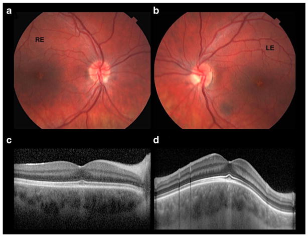
Fig. 2.
Patient 2: ataxia associated with hypogonadotropic hypogonadism—ophthalmological exam. a, b Color fundus photographs reveals a small depigmented foveal symmetrical spot in OU. c, d OCT horizontal scan of RE and OCT vertical scan in LE show foveal area with disruption of C.O.S.T (cone outer segment tips) and choroidal hyperreflectivity, demonstrating retinal pigment epithelium (RPE) atrophy at this site. RE right eye, LE left eye

