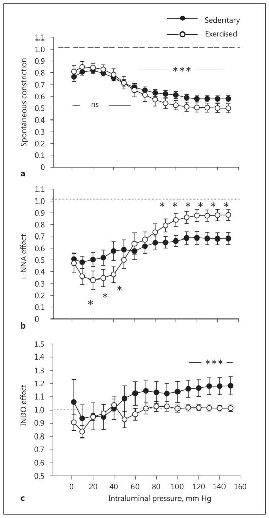
Fig. 3.
Exercise training-induced remodeling of rat intramural coronary arterioles compared with sedentary controls. a Inner diameter in spontaneous tone and myogenic constriction expressed as a ratio of the passive diameter. The dashed line refers to the diameter of arterioles in the passive condition, taken as the reference. Alteration of the spontaneously contracted inner diameter in response to the NOS inhibitor Nω-nitro-L-arginine (L-NNA, 10−5 M; b) and the cyclo-oxygenase inhibitor (INDO, 2.8 × 10⁻⁵ M; c). The dotted line (b, c), representing the diameter of arterioles in control myogenic constriction, was taken as a reference at each pressure level. Mean values were used from 2-way ANOVA tests with Tukey paired comparisons: * p < 0.05 and *** p < 0.001 between sedentary (n = 10) and exercised (n = 6) groups.

