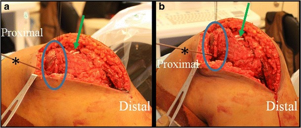
Fig. 6.

Intraoperative pictures of a MPFL reconstruction demonstrating isometry testing following placement of the guidewire (asterisk). Once the graft (blue circle) is looped around the femoral guidewire, the knee is taken through a range of motion to ensure that graft tension does not change. In these images, the two tails of the graft (green arrow) can be seen prior to their insertion on the patellar. Note that the MPFL was combined with additional procedures and thus the larger incision
