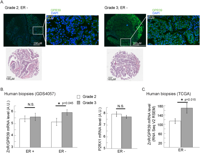
Figure 4.
ZnR/GPR39 expression is increased in higher grade breast cancer tissue. (A) An array of breast cancer biopsies (http://www.biomax.us/tissue-arrays/Breast/BR1504a) was stained for ZnR/GPR39. Average staining level, above a threshold, in ER negative samples of grade 2 and grade 3 tissues is shown (scale bar shown on image). Representative images of ZnR/GPR39 staining and hematoxylin eosin stain (from the company site) are shown (bottom panel). (B) Analysis of ZnR/GPR39 mRNA level in ER-positive or negative, HER2-normal, breast cancer grade 2 (white) or 3 (grey) tumors (GDS4057 cohort, left panel). Note that a significant increase is seen in ZnR/GPR39 expression in ER-negative grade 3 tumors (p < 0.05 t-test). Right panel shows the analysis of mRNA level of P2XR1 purinergic receptor on the same cohort. (C) Analysis of ZnR/GPR39 mRNA level in ER-positive or negative, breast cancer stage 2 (white) or 3 (grey) tumors (TCGA cohort, p < 0.05, Welch’s t-test).

