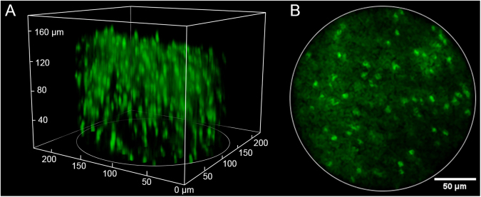
Figure 3.
(A) Two photon image showing a 3D volume acquired by the 2P-FCM (220 × 220 μm lateral × 180 μm axial) with over 200 cells in the image. The sample is fixed mouse cortical tissue (4% paraformaldehyde) expressing green fluorescent protein (GFP) driven by proteolipid protein promoter. The GFP labels oligodendrocyte cell bodies in the cortex. (B) Processed image of a single slice in the stack after filtering to remove pixelation pattern.

