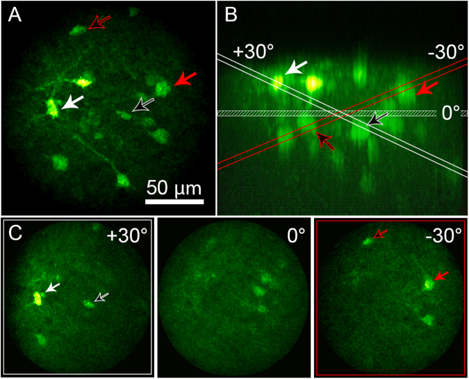
Figure 4.
Tilted-field scan enabled by rapid focusing of the EWTL lens. (A) Maximum intensity projection of a thick coronal section of mouse brain, expressing GCaMP6s in neurons, acquired by the 2P-FCM. Arrows indicate cell bodies that are visible during tilted-field scanning. (B) Side (XZ) projection of the image volume in A. The same cell bodies are indicated by the arrows. The planes for the horizontal and angled scan limits are indicated and color coded red for the −30 degrees scan, and white for the +30 degrees scan. (C) Images showing the tilted-field scans, indicating the same cell bodies that are shown to intersect with the red or white planes in B.

