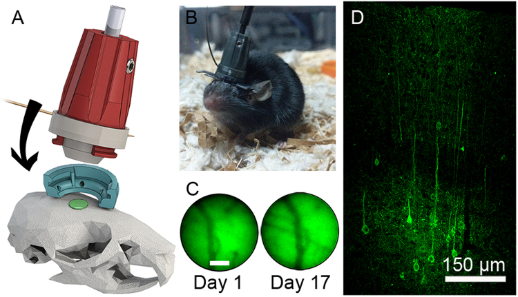
Figure 6.
Mouse implant and verification of GCaMP6s expression. (A) Attachment mechanism of 2P-FCM onto baseplate attached to the mouse skull. (B) Photo of a behaving mouse with 2P-FCM attached. (C) Verification of stability of imaging FOV over 17 days, showing the same blood vessels with epifluorescence measured through the FCM. (D) Confocal image of the neocortex for a fixed cortical coronal slice of the mouse’s brain 12 weeks post-injection of AAV5 expressing GCaMP6s driven under control of the synapsin promoter.

