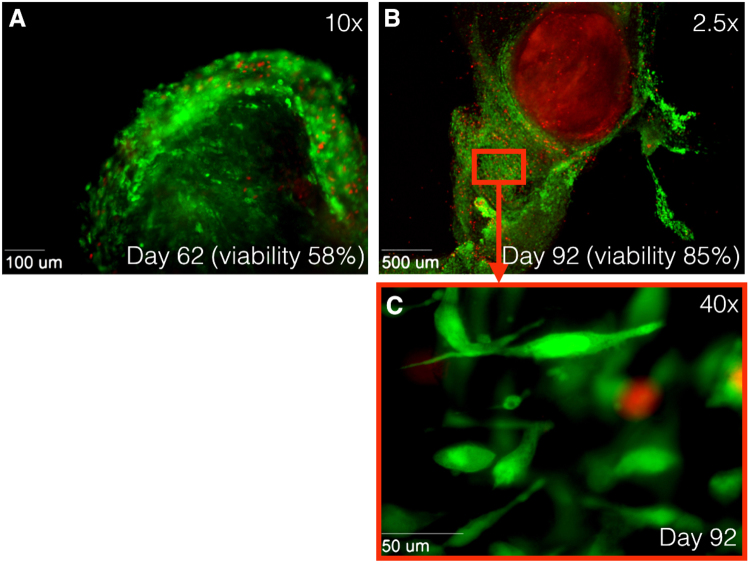
Figure 3.
Immunofluorescence images at 10× magnification using Zeiss Axiovert 200 M Marianas™ Microscope. Human tissue stained with LIVE/DEAD® Viability/Cytotoxicity Kit. Green fluorescence shows live cells, while red fluorescence indicates dead nuclei. (A) Alive tissue at day 62 after harvesting. Tissue viability of 58%. (B) Alive tissue at day 62 after harvesting at 2.5× magnification and (C) at 40× magnification. Outgrowth of new cells is observed after 92 days, while original tissue shows only staining of EthD-1 (dead cells). Tissue viability of 85%. EthD-1 indicates ethdium homodimer-1.

