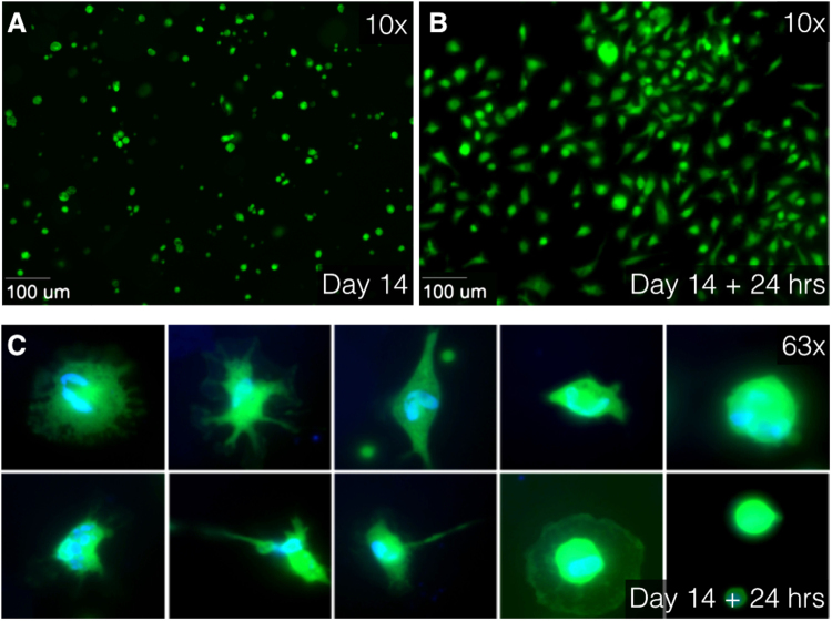
Figure 7.
Immunofluorescence images at 10× (A,B) and 63× oil (C) magnification using Zeiss Axiovert 200 M Marianas™ Microscope. (A) After 14 days of culturing, tissues were enzymatically digested using collagenase. Live separate cells, stained with Calcein AM (green), float as round cells in culture medium. (B) After 24 hours of additional culturing, the cells were attached to the slide. (C) Different cell type characteristics and differences in cell nuclei are observed in attached cells. Live cytoplasm is stained with Calcein AM (green) and cell nuclei are stained with DAPI (blue). AM indicates acetoxymethyl and DAPI, 4′,6-diamidino-2-phenylindole.

