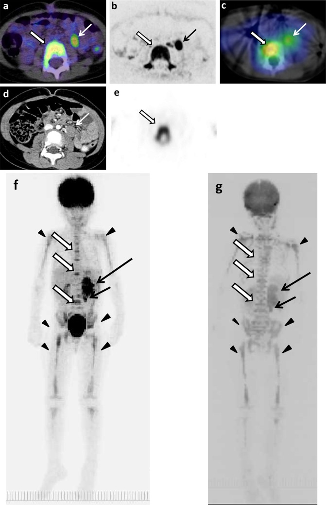
Fig. 3.
The original lesions in left adrenal gland, metastases in paraaortic lymph nodes and thoracic and lumbar spines, and false-positive images of skeletons of 4 years and 9 months old girl (patient 11). a 18F-FDG PET/CT. b DWIBS. c 123I-MIBG scintigraphy/SPECT-CT. d CT. e Bone scintigraphy/SPECT. The MIP of 18F-FDG PET (f) and DWIBS (g). Long arrows show the original lesion (f, g). Short arrows show lymph node metastases (a–g). Empty arrows show bone metastases (a–c, e–g). Arrowheads show false-positive images of various bone segments (f, g)

