Fig. 4.
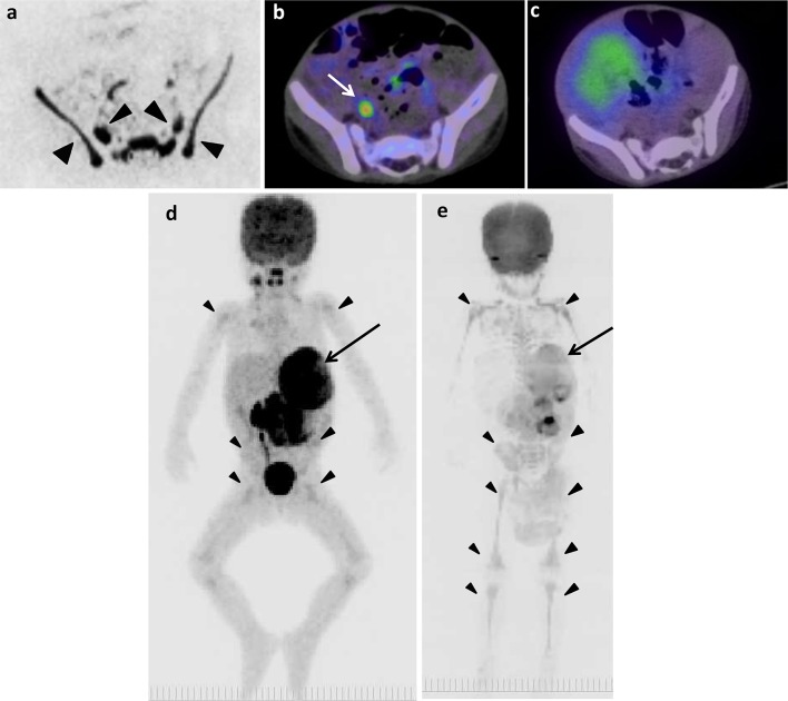
The original lesion in left adrenal gland and false-positive images of skeletons of 2 years and 2 months old boy (patient 8). a DWIBS. b 18F-FDG PET/CT. c 123I-MIBG scintigraphy/SPECT-CT. The MIP of 18F-FDG PET (d) and DWIBS (e). Long arrows show the original lesion (d, e). Arrowheads show false-positive images in pelvic bones (a) and various bone segments (d, e). Short arrow shows 18F-FDG uptake in right ureter (b)

