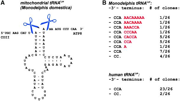
Figure 4.
(A) Secondary structure drawing of mitochondrial tRNALys from M. domestica within the precursor transcript. Scissors indicate 5′- and 3′-processing cleavage positions as determined from cDNA clone analysis after circularization or tagging of the tRNA. (B) Posttranscriptional addition of the CCA terminus and further 3′-terminal extensions (shown in red). Numbers indicate the frequency at which individual clones were observed.

