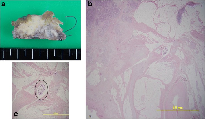
Fig. 2.
a A cut surface of the pancreatic body showed a fibrotic tissue in the area where the tumor was located (HF-1-05). b On histological analysis, 99% of the cancer cells had disappeared and had been replaced with fibrotic tissue. c High-power photomicrograph revealed a minute residual cancer tissue (circle)

