Fig. 3.
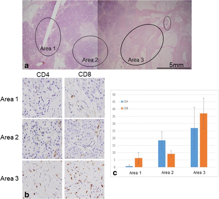
Evaluation of CD4+ and CD8+ cells infiltration around the cancer tissue (HF-1-05). a Three areas in different distances (circle) from the residual cancer (dot-line circle) were evaluated. b, c Infiltration of CD4+ and CD8+ cells was significant in the fibrosis near the residual cancer tissue (area 3) and it became obscure as the areas receded from the cancer tissue

