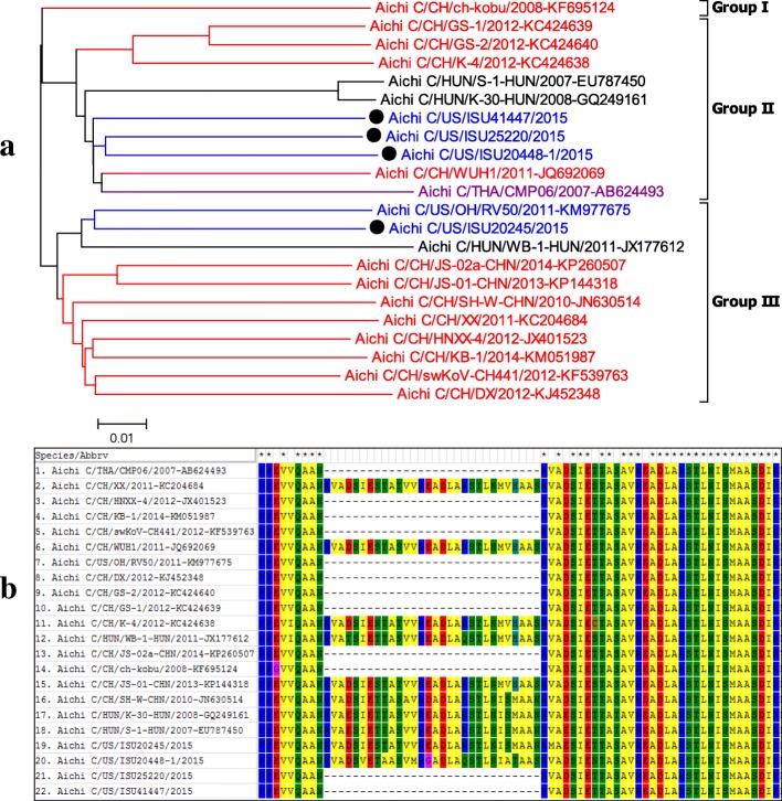
Fig. 5.
a, phylogenetic tree analysis of polyprotein of different Aichivirus C (PKV-1) strains. Based on phylogenetic analysis with whole genome sequences, kobuviruses form three genogroups. The kobuviruses detected in this study (Black dot) were categorized into two different genogroups. One strain (ISU20245) is most closely related to a US strain reported in 2011, and three additional strains (ISU20448, ISU25220, and ISU41447) were grouped into to anther genogroup and were most closely related to Chinese and Thai strains implicated in swine diarrhea. b, Aichivirus C (PKV-1) alignment (aa position 1183–1224 of polyprotein with PKV/US/ISU25220/2015 as reference) demonstrating insertions and deletions among different strains

