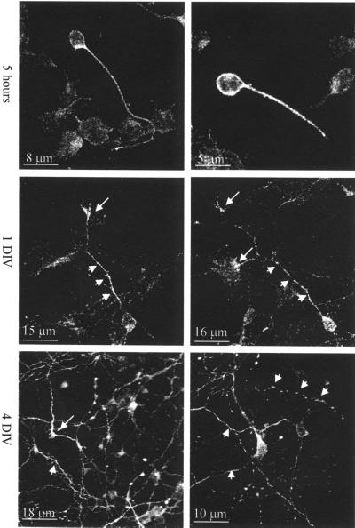
Figure 2.
Immunofluorescence analysis of palladin during development of primary cortical neurons. Neurons cultured for 5 h, 1 d, and 4 d were fixed with 4% paraformaldehyde, permeabilized with 0.2% Triton X-100, and then stained for palladin. After 5 h in culture, palladin is localized in the cell body and the first process. In 1-DIV neurons, palladin localized to the cell body and the nascent axon (short arrows) and axonal growth cone (long arrows). In 4-DIV neurons, palladin is concentrated in the processes (short arrows) and axonal growth cones (long arrows).

