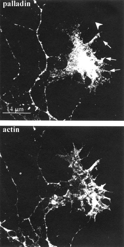
Figure 5.
Palladin and F-actin localization in the axonal growth cone. Cultured cortical neurons (4 DIV) were fixed, permeabilized, and then double-stained for palladin and filamentous actin. Palladin is concentrated in the central region of the growth cone and partially colocalized with F-actin in some, but not all, of the filopodia. Arrows point to filopodia that stain positively for palladin; single arrowhead points to a filopodium that failed to stain for palladin.

