Figure 1.
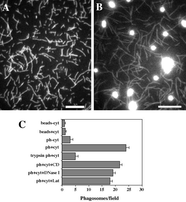
Reconstitution of salt-stripped phagosome binding to preassembled F-actin in vitro. (A and B) Fluorescence images of typical fields of the binding assay. (A) Rhodamine-phalloidin–labeled F-actin was absorbed on the coverslip surface. (B) Phagosomes were bound to F-actin in the presence of 1 mg/ml cytosol and unbound phagosomes were washed out. Bars, 10 μm. (C) Stimulation of phagosome binding to F-actin by cytosolic (cyt) factor(s). Binding to the F-actin lawn of uninternalized latex beads (beads−cyt; beads+cyt). Phagosome (ph) binding to F-actin in the presence and absence of 1 mg/ml cytosol (ph+cyt, ph−cyt). Binding of phagosomes treated with 100 μg/ml trypsin (trypsin ph+cyt) (see MATERIALS AND METHODS). Binding of phagosomes in the presence of 5 μM cytochalasin D (ph+cyt+CD), 15 μM DNase I (ph+cyt+DNase I), or 5 μM latrunculin (ph+cyt+Lat).

