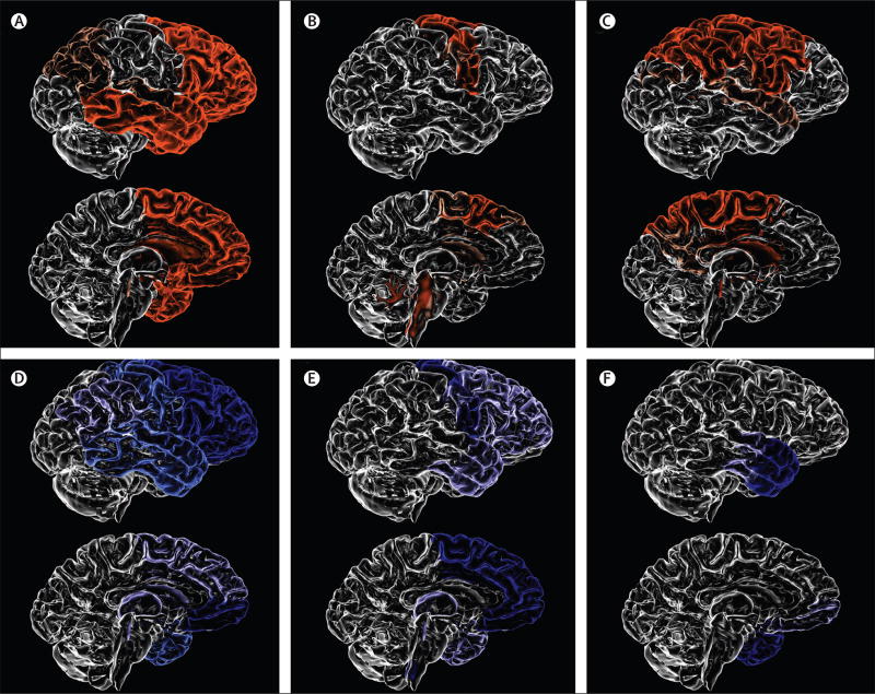
Figure 3. Patterns of brain atrophy in FTLD pathologies.
(A) Pick’s disease; (B) progressive supranuclear palsy; (C) corticobasal degeneration; (D) FTLD-TDP type A; (E) FTLD-TDP type B; (F) FTLD-TDP type C. FTLD-TDP type A is associated with an asymmetrical dorsal pattern of atrophy that includes the frontal lobe and temporal lobe (anterior, medial, and posterior regions), orbitofrontal cortex, anterior cingulate gyrus, inferior parietal lobe, striatum, and thalamus. FTLD-TDP type B is associated with a more medial pattern of atrophy mainly involving the medial and polar temporal lobe, anterior insular, cingulate and medial prefrontal cortices, and the orbitofrontal cortex. The frontal lobe is more severely affected in the posterior areas. FTLD-TDP type C is associated with either right-predominant or left-predominant anterior temporal lobe atrophy, with additional involvement of the amygdala, hippocampus, orbitofrontal cortex, and insular cortex. FTLD=frontotemporal lobar degeneration. TDP=TAR DN A-binding protein 43.

