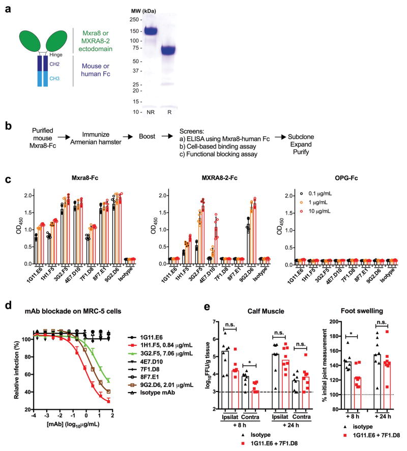
Extended Data Figure 8. Mxra8-Fc and anti-Mxra8 generation and function.
a. Diagram of Mxra8-Fc (left) and SDS-PAGE (non-reducing [NR] and reducing [R] conditions) of purified material (right). Data are representative of three experiments. b. Scheme of anti-Mxra8 generation in Armenian hamsters. c. ELISA reactivity of anti-Mxra8 mAbs against Mxra8-Fc, MXRA8-2-Fc, or OPG-Fc. Purified proteins (50 μl, 5 μg/ml) were immobilized overnight at 4°C on ELISA plates. Anti-Mxra8 and isotype control mAbs were incubated for 1 h at room temperature. Signal was detected at 450 nm after incubation with horseradish peroxide conjugated goat anti-Armenian hamster IgG (H+L) and development with 3,3′-5,5′ tetramethylbenzidine substrate. d. Blockade of CHIKV-181/25 infection in MRC-5 cells with seven different hamster anti-Mxra8 or isotype control mAbs. MAbs were pre-incubated with cells for 1 h at 37°C prior to addition of virus. After infection, cells were processed for E2 staining by flow cytometry. Relative infection was compared to a no mAb condition using flow cytometry and anti-E2 staining. Data in c and d are pooled from two experiments (n = 6) and expressed as mean ± SD. e. Anti-Mxra8 mAbs (1G11 + 7F1) or isotype control hamster mAbs (300 μg total) were administered via intraperitoneal route 8 or 24 hours after inoculation of CHIKV-AF15561 in the footpad. (Left) At 72 h after initial infection, CHIKV titers were measured in the ipsilateral and contralateral gastrocnemius (calf) muscles. (Right) At 72 h, ipsilateral foot swelling was measured and compared to measurements taken immediately prior to infection. Data are pooled from two experiments (n = 8; *, P < 0.05; two-tailed Mann-Whitney test) and expressed as median values.

