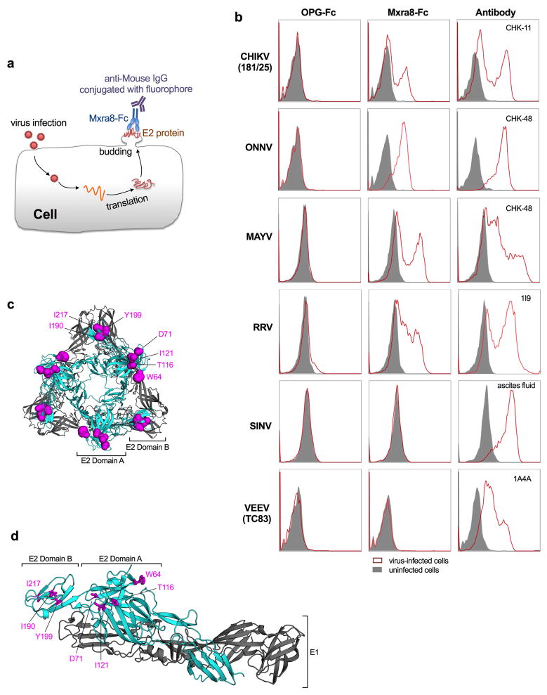
Extended Data Figure 10. Binding of Mxra8-Fc to surface-displayed E2 protein in virus-infected cells.
a. Diagram of the cell-based binding assay. After infection, viral structural proteins (e.g., E2) traffic to the cell plasma membrane where progeny virion assembles and buds. E2 protein is displayed on the cell surface and is accessible to the binding of Mxra8-Fc and detection with a goat anti-mouse IgG secondary antibody by flow cytometry. b. Binding of Mxra8-Fc to virus-infected WT 3T3 cells. Cell were infected with the indicated viruses and processed for Mxra8-Fc binding by flow cytometry. Virus-specific anti-E2 Abs were used as positive controls. Data are representative of two independent experiments. c–d. Mapped residues by are shown as magenta spheres (c) or sticks (d) on the CHIKV p62-E1 structure (c, trimer of dimers, top view; d, heterodimer, side view) using PyMOL (PDB 3N41). The E1 and E2 proteins are colored in grey and cyan, respectively.

