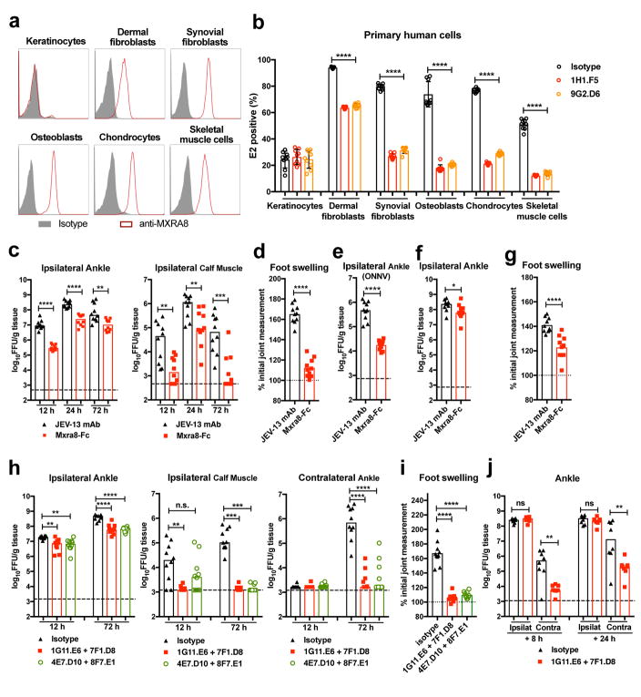
Figure 4. Mxra8 contributes to alphavirus pathogenesis.
(a) Surface expression of MXRA8 on primary human keratinocytes, dermal fibroblasts, synovial fibroblasts, osteoblasts, chondrocytes, and skeletal muscle cells. One experiment of three is shown. (b) Cells were pre-incubated with anti-MXRA8 blocking mAbs prior to addition of CHIKV-AF15561 and processed for E2 staining (3 experiments, n = 9; one-way ANOVA with Dunnett’s test, ****, P < 0.0001). c–d. Mxra8-Fc or JEV-13 mAb were incubated with CHIKV-AF15561 for 30 min prior to subcutaneous inoculation. (c) At 12, 24, and 72 h, CHIKV was measured in the ankle and calf muscle. (d) At 72 h, foot swelling was measured (2 experiments, n = 10; median viral titers: *, P < 0.05; **, P < 0.01; two-tailed Mann-Whitney test; mean foot swelling, ****, P < 0.0001; two-tailed unpaired t-test). e. Mxra8-Fc or JEV-13 mAb were mixed with ONNV immediately prior to subcutaneous inoculation. At 12 h, ONNV was measured in the ankle (2 experiments, n = 10; ****, P < 0.0001; two-tailed unpaired t-test; median values). f–g. Mxra8-Fc or JEV-13 mAb were administered via an intraperitoneal route 6 h prior to CHIKV-AF15561 inoculation in the footpad. At 24 h, CHIKV was measured in the ankle (f). At 72 h, foot swelling was measured (g) (2 experiments, n = 10; median viral titers: *, P < 0.05; two-tailed Mann-Whitney test; mean foot swelling, ****, P < 0.0001; two-tailed unpaired t-test). h–j. Pairs of anti-Mxra8 mAbs or isotype control hamster mAbs were administered via an intraperitoneal route 12 h prior to (h–i) or 8 or 24 hours after (j) inoculation of CHIKV-AF15561. At 12 (h) and 72 (h, j) h, CHIKV was measured. At 72 h, foot swelling (i) was measured (2 experiments, (h (left) and i: n = 10; **, P < 0.01; ****, P < 0.0001; one-way ANOVA with Dunnett’s test; h (middle and right): n = 10; **, P < 0.01; ***, P < 0.001; ****, P < 0.0001; Kruskal-Wallis with Dunn’s test; j: n = 8; **, P < 0.01; two-tailed Mann-Whitney test).

