. 2018 May 3;15(3):213–223. doi: 10.21873/cgp.20080

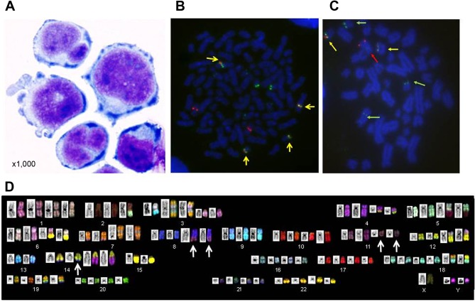
Figure 1. Morphological and cytogenetic features of KPUM-YY1 cells. A: Morphology of KPUM-YY1 cells. Cells were cytospinned and stained using Diff-Quik (Sysmex, Kobe, Japan), and the morphology was examined under a light microscope (magnification ×1,000). B: Metaphase double color (DC)-fluorescence in situ hybridization (FISH) analysis for immunoglobulin heavy chain (IgH) and cyclin D1 (CCND1) in KPUM-YY1 cells. Red and green signals indicate CCND1 at 11q13 and IgH at 14q32, respectively. Yellow signals (arrows) indicate fusion of red and green signals, confirming the reciprocal translocation IgH/CCND1. C: Metaphase DC-FISH analysis for IgH and c-MYC in KPUM-YY1 cells. Red and green signals indicate c-MYC at 8q24 (red arrows) and IgH at 14q32 (green arrows), respectively. Yellow signals (arrows) indicate fusion of red and green signals, confirming the reciprocal translocation IgH/c-MYC. D: SKY analysis of KPUM-YY1 cells. The representative karyotype of KPUM-YY1 is 4n: 88, YY, inv(X) (p11q28), t(2;5)(q21;q15), -2,del(3)(p12)x2,+der(3)t(3;13)(q12;q14), der(3)(7qter→7q22::3p21 →3q21::13::7q11.2→7qter)x2, t(4;14)(q12;q32.1)x2, der(5)t(2;5), der(6)t(6;8)(p25;q24)x2, der(6)t(6;15)(q13;q11)x2, add(7)(q22)x2, der(7)(7qter→7q11.2::8::7p22→7q11.2::7p22→7pter)x2, t(8;14;11)(q24;q32;q13)x2, dup(9)(p13p24)x3, -9, der(10)t(10;19) (p15;q13) x2, t(12;18)(q15;p11.2)x2, der(12)t(3;12)(p21;p11.2)x2, -13x2, -15x2, +18x2, der(19)(19pter→19q13.1::14q24→14q32:: 8q24→8qter)x2. Arrows indicate a three-way translocation t(8;14;11)(q24;q32;q13).

Figure 1