. 2018 May 3;15(3):213–223. doi: 10.21873/cgp.20080

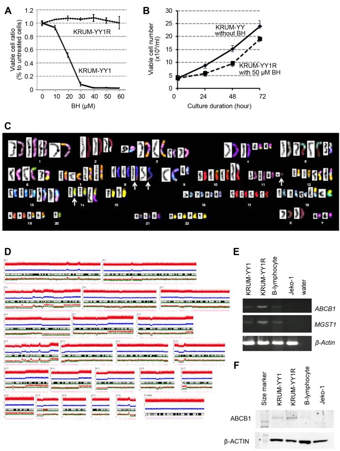
Figure 2. Cellular and cytogenetic/molecular characteristics of bendamustine (BH)-resistant KPUM-YY1R cells. A: Growth inhibitory effect of BH on KPUM-YY1 and KPUM-YY1R cells. Cells were treated with BH for 96 h and cell viability was evaluated by a modified MTT assay. Data are shown as means+/-standard errors from triplicate experiments. Growth of KPUM-YY1 cells was inhibited by BH in a dosedependent manner, while growth of KPUM-YY1R cells was not inhibited by 60 μM BH. B: Growth curves of KPUM-YY1 and KPUM-YY1R cells. KPUM-YY1 cells were cultured in normal medium, while KPUM-YY1R cells were grown in culture medium containing 50 μM BH. Cells were stained by Trypan Blue and viable cell numbers were counted under an inverted microscope. C: SKY analysis of KPUM-YY1R cells. The representative karyotype of KPUM-YY1R cells is 4n: 84, YY, inv(X)(p11q28)x2, der(1)t(1;7)(p32;q11.2), t(2;5)(q21;q15), -2, del(3)(p12)x2, +der(3)t(3;13)(q12;q14), der(3)(7qter→7q22::3p21→3q21 ::13::7q11.2→7qter)x2, t(4;14)(q12;q32.1), der(4)t(4;14), der(5)t(2;5), der(5)t(5;9)(q11;p13), der(6)t(6;15)(q13;q11), der(6)t(6;8)(p25;q24)x2, dic(6;20)(p21;q13), -6, add(7)(q22)x2, der(7)(7qter→7q11.2::8::7p22→ 7q11.2::7p22→7pter)x2, t(8;14;11)(q24;q32;q13)x2, del(9)(q21), der(9)(9pter→9q22::13::5), -9, -9, der(10)t(10;19)(p15;q13)x2, der(10) (5qter→5q15::2q21→2q13::10p11→10qter), t(12;18)(q15;p11.2)x2, der(12)t(3;12)(p21;p11.2)x2, der(13)t(1;13)(p22;q32)x2, +13, -14, -15, - 15, +18, der(19)(19pter→19q13.1::14q24→14q32:: 8q24→8qter)x2, -20, -20, -20, -21. Arrows indicate a three-way translocation t(8;14;11)(q24;q32;q13). D: Genome copy number analysis in KPUMYY1R cells. A DNA gain and loss assay based on a SNP array was performed on genomic DNA purified from KPUM-YY1R cells. The CNAG3.5.1 program was used for analysis of SNP array data to determine total copy numbers. E: RT-PCR analyses for ABCB1 and MGST1. Transcriptional expression of ABCB1 and MGST1 was examined in KPUM-YY1 cells, KPUM-YY1R cells, normal B lymphocytes, and Jeko-1 cells (MCL-derived cell line). β-Actin was used as an internal control. F: Western blot for ABCB1 protein. ABCB1 protein expression was examined in KPUM-YY1 cells, KPUM-YY1R cells, normal B lymphocytes, and Jeko-1 cells, with β-Actin used as an internal control.

Figure 2