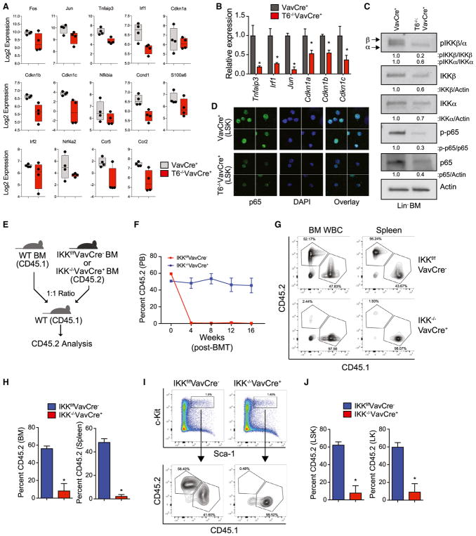
Figure 6. Reduced NF-κB Signaling and IKKβ Deficiency Leads to Loss of HSPC and Impaired Hematopoietic Reconstitution.
(A) Expression of NF-κB target genes in VavCre+ and Traf6−/−VavCre+ LSK cells as determined by RNA-seq.
(B) Expression of select NF-κB target genes measured by qRT-PCR in Traf6−/−VavCre+ relative to VavCre+ LSK cells.
(C) Immunoblot analysis of phosphorylated IKKβ, IKKα, and p65 in VavCre+ and Traf6−/−VavCre+ Lin− BM cells. Shown below is the relative expression of the indicated proteins calculated by measuring densitometry of the immunoblots.
(D) Expression and localization of p65 (green) in VavCre+ and Traf6−/−VavCre+ LSK cells as determined by indirect immunofluorescence (IF). Shown are representative images from 3 pooled mice per group.
(E) Outline of competitive BM transplantations using IKKf/fVavCre− or IKK−/−VavCre+ mice.
(F) Summary of donor-derived PB proportions at the indicated time points after competitive transplantation using IKKf/fVavCre− or IKK−/−VavCre+ BM cells (n = 5 per group). Two independent experiments were performed.
(G) Representative flow cytometric analysis of donor-derived (CD45.2+) and competitor-derived (CD45.1+) BM and spleen cells from recipient mice after competitive transplantation using IKKf/fVavCre− or IKK−/−VavCre+ BM cells.
(H) Proportion of donor-derived IKKf/fVavCre− or IKK−/−VavCre+ BM and spleen cells 8 weeks after competitive BM transplantation (n = 5 per group).
(I) Representative flow cytometric analysis of donor-derived (CD45.2+) and competitor-derived (CD45.1+) BM LSK cells from recipient mice after competitive transplantation using IKKf/fVavCre− or IKK−/−VavCre+ BM cells.
(J) Proportion of donor-derived IKKf/fVavCre− or IKK−/−VavCre+ LSK BM 8 weeks after competitive BM transplantation (n = 5 per group). *p < 0.05. Data are represented as mean ± SEM.

