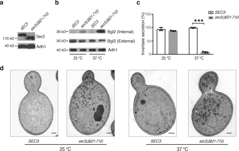
Figure 6. The Sec3 CorEx deletion mutant is defective in exocytosis.
a. Expression of Sec3-GFP and Sec3(Δ621–710)-GFP in yeast cell lysates was detected by Western blotting with an anti-GFP antibody. Alcohol dehydrogenase-1 (“Adh1”) was used as a loading control. b. Accumulation of Bgl2 in sec3(Δ621–710) cells at 25°C and 37°C. Internal and external Bgl2 was detected by Western blotting. Adh1 was used as a loading control. Uncropped blot images are shown in Supplementary Data Set 4. This experiment was independently performed twice with similar results. c. Accumulation of invertase in sec3(Δ621–710) cells at 37°C. The mean and s.d. value of the invertase secretion rate was shown as bars and error bars, respectively. n=3, as technical replicates (see METHOD); “***”, P<0.001. This experiment was independently performed twice with similar results. d. Accumulation of post-Golgi secretory vesicles in sec3(Δ621–710) cells as revealed by thin-section EM. Scale bar, 0.5 µm. At least five images were taken for each condition, showing similar results.

