
Figure 2.
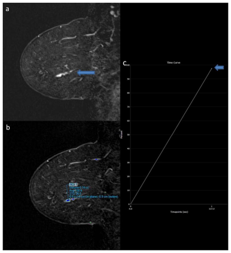
58-year-old woman with history of bilateral papillomas status post surgical excision. New 2.0 cm linear clumped nonmass enhancement is present on the sagittal T1-weighted post-contrast subtraction image of the left breast on surveillance MRI (2A, arrow). Lesion ROI (2B) demonstrated IER of 98% when measured by Reader 1 (2C, arrow) and 72% by Reader 2 on first post-contrast images. MR-guided biopsy yielded intermediate-grade ductal carcinoma in situ.