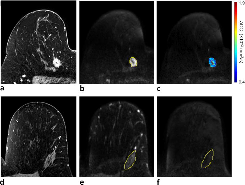
Figure 4.
Example of invasive lobular carcinoma (grade 2/3) with low RS (RS = 17) detected in a 33-year-old female. a) DCE-MRI demonstrates a 47 mm right breast mass at 3 o’clock, 60 mm from the nipple. On DWI, the lesion exhibits b) relatively low signal intensity on b = 800 s/mm2 diffusion-weighted image with CNR = 1.1 (lesion ROI contour is shown), and c) moderate to low ADC (ADCmean = 1.15 × 10⁻³ mm2/s). For CNR calculation, d) normal fibroglandular tissue was identified on DCE-MRI at a corresponding slice level in the contralateral breast, e) an ROI was defined on the b = 0 s/mm2 image, where breast tissue is most visible, to cover the largest tissue area possible, f) which was then propagated to the b = 800 s/mm2 image to measure the mean DWI signal intensity within the ROI.

