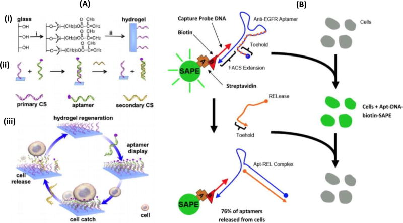
Figure 3.
(A) Aptamer-mediated CTC capture and release via complementary oligonucleotide sequences (CS= complementary sequences) (Zhang et al., 2012). (i) explain about the basic structure of the of the hydrogel immobilized on the glass slide (ii) illustration of the aptamer sequence hybridized with complementary sequences (CSs) (iii) represents the process of cell release. The stable aptamer hybridized with triggering CSs, forming a new hybridized state that leads to rapid dissociation of the aptamer from the hydrogel hence releasing the cells from the hydrogel. (B) Illustrating the cell binding and aptamer formed by SAPE of anti-EGFR aptamer – DNA and biotin, and addition of RELease particles. After washing, the fluorescence cells (caused by selective binding with aptamer) completely hybridizes with anti-EGFR aptamer because of the presences of RELease RNA. This leads to opening up its herpin sturucture which releases 76% of anti-EGFR aptamer from the cellular surface. (Wan et al., 2012)

