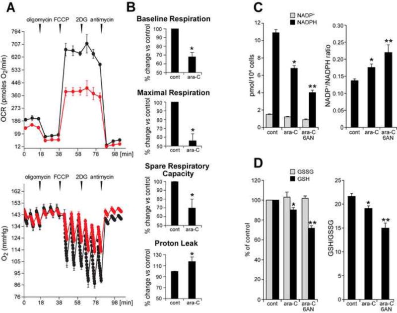
Figure 5. Ara-C exposure compromises mitochondrial respiration and decreases NADPH and GSH levels in DRG neurons.
(A) Respiratory profile for non-treated control DRG neurons (black) shows baseline OCR of ~180 pmoles O2/min. Sequential, in port additions of mitochondrial effectors (vertical arrows), reveal that ~90% of total oxygen consumption feed mitochondrial respiration: of this OCR ~70% support ATP synthesis and ~30% is lost to proton leak. Spare respiratory capacity (SRC), i.e., FCCP-induced increase in OCR over the baseline is ~250% for control DRGs. Following ara-C exposure baseline OCR (red) drops by ~30% and maximal respiration by nearly 50% (FCCP-induced), with ~30% reduction in SRC and ~ 20% increase in OCR feeding the proton leak. (Bottom) Oxygen tension in control wells is reduced by the addition of FCCP and even further by 2-deoxyglucose (2DG). Oxygen tension is reduced to a lesser extent in ara-C exposed DRGs, reflecting diminished mitochondrial OCR. (B) Each parameter calculated for control cultures was assigned the value of 100%; effects of treatment were calculated as percent change relative to each respective control. Data are presented as mean±SEM of four independent biological experiments. (C) NADPH levels are decreased and NADP+/NADPH ratios increased by ara-C. Concomitant addition of 6AN increased these effects. Data are mean±SEM for 3 independent experiments; * different from control; **different from ara-C; P< 0.05. (D) GSH levels were reduced by ara-C and further depleted by 6AN. Data are given as mean±SEM calculated from 3 independent experiments; * different from control; **different from ara-C; P< 0.05

