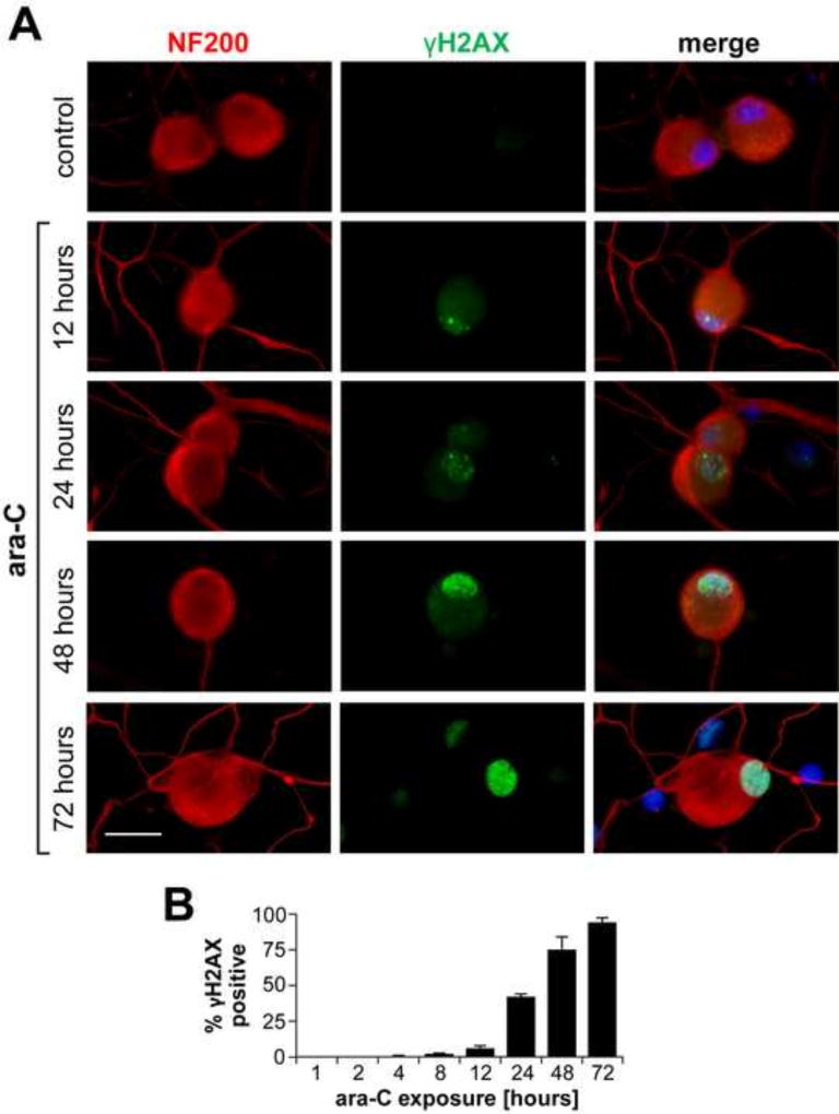
Figure 6. Temporal induction of nuclear γH2AX foci in ara-C exposed DRG neurons.
Representative images of time dependent formation of nuclear γH2AX foci during ara-C exposure of DRG neurons are shown (A). DRG neurons are identified by NF200 (red) and γH2AX foci are observed in green; sparse foci emerge by 12 h with density increasing in the course of 72 h ara-C exposure (scale bar = 20 µm). B) Percent of γH2AX positive nuclei increased over time with positive DRGs reaching ~35%, 75% and 95% by 24 h, 48 h and 72 h, respectively. Data are obtained from three independent biological experiments and presented as mean±SEM percent of γH2AX positive nuclei versus time.

