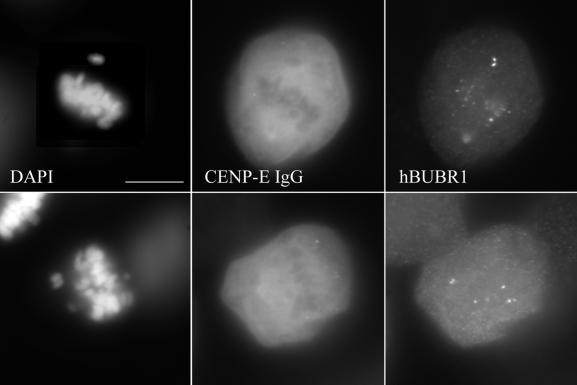
Figure 10.
Localization of hBUBR1 in anti-CENP-E–injected CF-PAC cells. Chromosomes were stained with DAPI (a and d) and injected HX-1 antibodies with Cy-5 anti-rabbit secondary antibody (b and e). hBUBR1 was detected with rat anti-hBUBR1 primary and Alexa Fluor 488 anti-rat secondary antibodies (c and f). Note the bright hBUBR1 staining of both sister kinetochores on the monooriented chromosomes. As in Figure 6, centromeres of injected cells show no HX-1 staining. Bar, 20 μm.

