Figure 7.
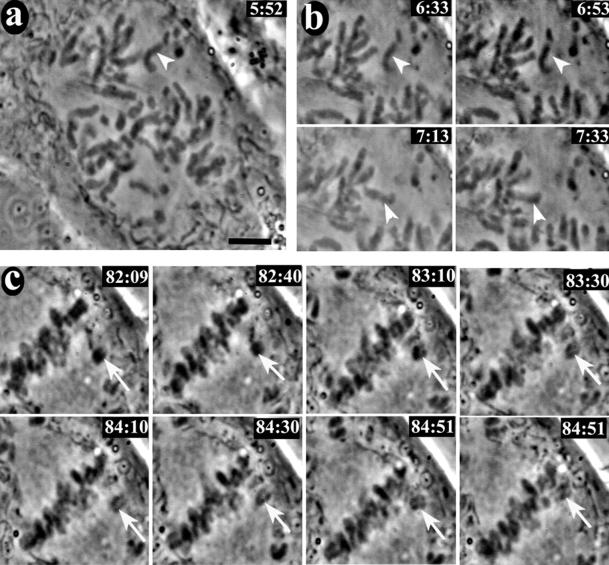
Video live cell imaging of a CF-PAC cell injected with HX-1 anti-CENP-E antibody. Synchronized CF-PAC cells were microinjected with antibody shortly after release from G1/S block and later remounted into Rose chambers. Coverslips were scanned for injected cells in prophase (injected cells were located via a scribe mark and coinjection with Oregon Green dextran). This cell was filmed for at total of 2 h past NEB with a 10-s filming rate (approximate time in minutes: seconds past NEB is located the upper right of each frame). (a) Early prometaphase. The unattached chromosome indicated by the arrowhead was about become monooriented and undergo fast poleward motion (see video 1 in online supplement). (b) Windowed frames showing the chromosome indicated in a undergoing fast motion to the upper spindle pole (see video 2 in online supplement). (c) Windowed frames illustrating congression. The congressing chromosome exhibited a typical transient reversal of motion before completing congression to the spindle equator (see video 3 in online supplement). Bar, 5.0 μm.

