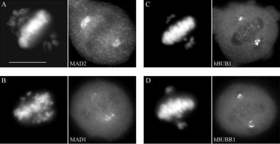
Figure 9.
Immunofluorescence staining of checkpoint proteins in anti-CENP-E–injected HeLa cells. Synchronized cells were injected with rat anti-CENP-E antibodies 2 h after release from the G1/S boundary and sampled 10 h later. (a–d) Cells were stained for hMAD2, hMAD1, hBUB1, and hBUBR1 with the use of appropriate rabbit antibodies and Texas Red-conjugated anti-rabbit secondary antibodies. Chromosomes and nuclei were stained with DAPI (left column). Injected cells exhibited the normal dissociation of checkpoint proteins from kinetochores of bioriented, aligned chromosomes. Bar, 10 μm.

