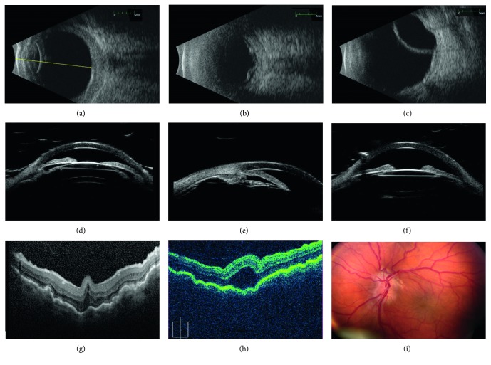
Figure 1.
Typical ultrasonographic and retinal features of nanophthalmos. (a–c) B-scan ultrasounds showing features of nanophthalmos including short axial length, thickened sclera, and choroid (a), serous retinal detachment (b), and choroidal effusion (c). (d–f) Ultrasound biomicroscopy in a nanophthalmic eyes showing shallow anterior chamber (d), angle closure (e), and anterior rotation of the lens-iris diaphragm (f). (g) Heidelberg Spectralis OCT showing prominent choroidal and retinal folds in a small eye. (h) Zeiss Cirrus OCT showing foveoschisis and choroidal folds in a nanophthalmic eye. (i) Fundus photos in a patient with nanophthalmos and optic disc drusen, showing chorioretinal folds and crowded disc with mild vascular tortuousity.

