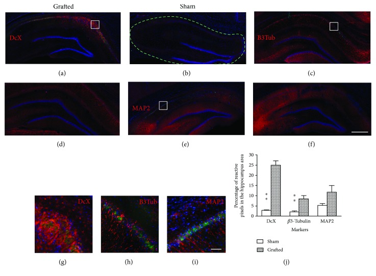
Figure 3.
Assessment of hippocampal neurogenesis after OE-MSC transplantation in ischemic rats. Immunohistological analyses revealed an increase of neurons in the hippocampus of grafted animals. (a) Newborn neurons expressing DcX (red) were mostly observed in the CA1 and CA2 areas of grafted animals, but none was observed in sham animals (b). Mature neurons expressing (red) β3-tubulin (c and d) or MAP2 (e and f) were also present in the hippocampi of animals from both groups. Higher magnification images revealed that no cells express both GFP (green) and tested neural markers (red) (g–i). The number of DcX and β3-tubulin-positive cells was significantly higher in the grafted group when compared to sham, and a tendency is observed for the MAP2 marker (p = 0.69) (j). Each image is representative of different animals from both groups. Scale bar: 1 mm (a–f), 100 μm (g–i). ∗∗p < 0.01. DcX: doublecortin; B3Tub: β3-tubulin; MAP2: microtubule-associated protein 2. Dashed line: selected area for antibody quantification.

