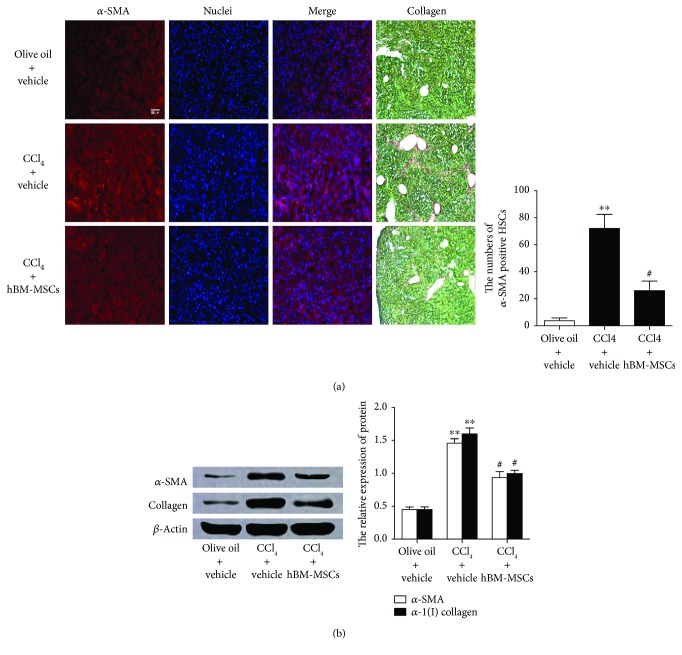
Figure 6.
hBM-MSCs lead to the decrease in liver fibrosis in the mouse model of CCI4-induced liver injury. Mice were divided into three groups and were, respectively, given administration of olive oil plus vehicle, CCI4 (5 μl/g body weight, two times a week) plus vehicle or CCI4 (5 μl/g body weight, two times a week) plus hBM-MSCs (8 × 10⁶/mouse). (a) Single fluorescence staining of α-SMA on the liver sections was detected (red fluorescence); the nuclei (blue fluorescence) were counterstained with Hoechst 33342. The images were captured with the fluorescence microscope. Scale bar = 100 μm. And stain collagen was examined by Sirius red staining of collagen. The representative images were captured with a light microscope. Scale bar = 50 μm. The number of α-SMA-positive HSCs in six randomly chosen fields was counted at 100-fold magnification, and the average values were shown. ∗∗P < 0.001 versus the control group. #P < 0.05 versus the mice of group treatment with CCI4 plus vehicle. (b) The protein levels of α-SMA and α1(I) collagen were examined by Western blot analysis after the HSCs were isolated from each group. ∗∗P < 0.001 versus the control group. #P < 0.05 versus the mice of group treatment with CCI4 plus vehicle.

