Figure 1.

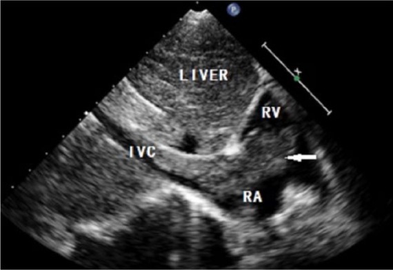
Echocardiography showing an isoechoic to hyperechoic mass (arrow) emerged from the IVC to the RA, passing through the tricuspid valve into the RV in diastole. RA: right atrium; RV: right ventricle; IVC: inferior vena cava.

Echocardiography showing an isoechoic to hyperechoic mass (arrow) emerged from the IVC to the RA, passing through the tricuspid valve into the RV in diastole. RA: right atrium; RV: right ventricle; IVC: inferior vena cava.