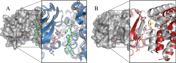
Figure 6.
Domain closure. A, surface view of P. maximus ODH (PmODH) “closed form” (PDB code 3C7C) showing the active site cleft with NADH in white carbons and the pentahistidine tag in green carbons. The expanded view shows a ribbon diagram of PmODH “open form” active site (blue) overlaid with the “closed form” (gray). Note the lack of conformational change indicating that both structures have open active sites. B, surface view of E. coli KPR (PDB code 2OFP) in a closed conformation with pantoate and NADPH bound. The expanded view shows a ribbon diagram overlay of KPR using the NADPH-binding domain for alignment. The holo form is in gray, and the apo form is in red (PDB code 1KS9). Ligands are NADPH (white) and pantoate (yellow). The dashed line is 3.3 Å. The holo form shows 24° closure of the catalytic domain as calculated by DynDom.

