
Figure 4.
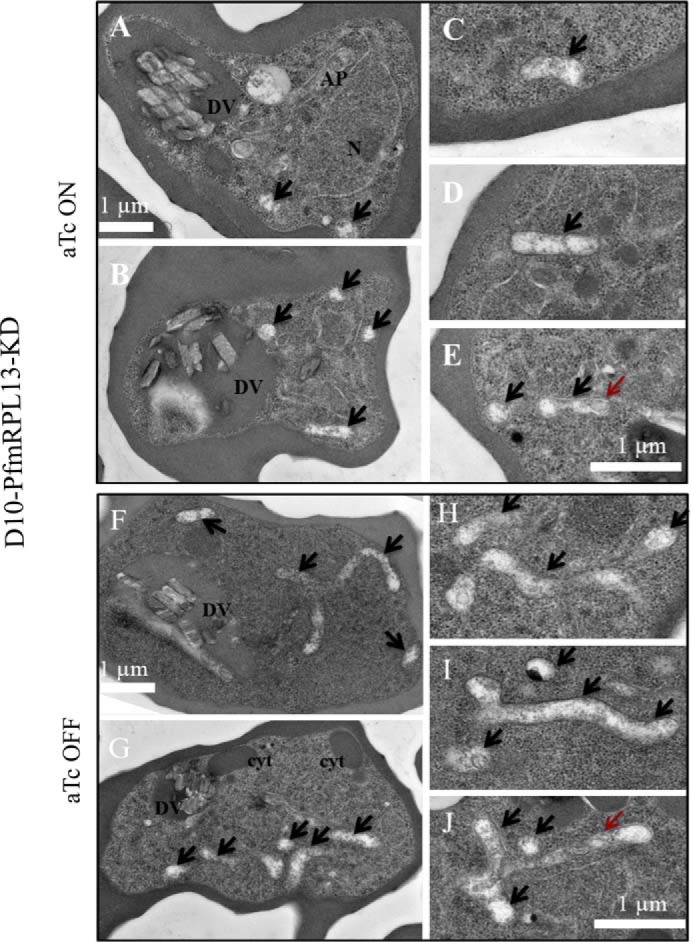
Morphological changes of mitochondria in the PfmRPL13 knockdown parasites maintained with or without aTc under transmission EM. A–E, TEM images of PfmRPL13 knockdown parasites maintained constantly under aTc. DV, digestive vacuole. AP, apicoplast. N, nucleus. F–J, TEM images of PfmRPL13 knockdown parasites grown without aTc for three cycles. DV, digestive vacuole. Cyt, cytostome. Black arrows indicate mitochondrial sections. Red arrows indicate internal membranes within the mitochondrial matrix. Each panel shows either an entire (A, B, F, G) or a partial (C–E, H–J) image of TEM.