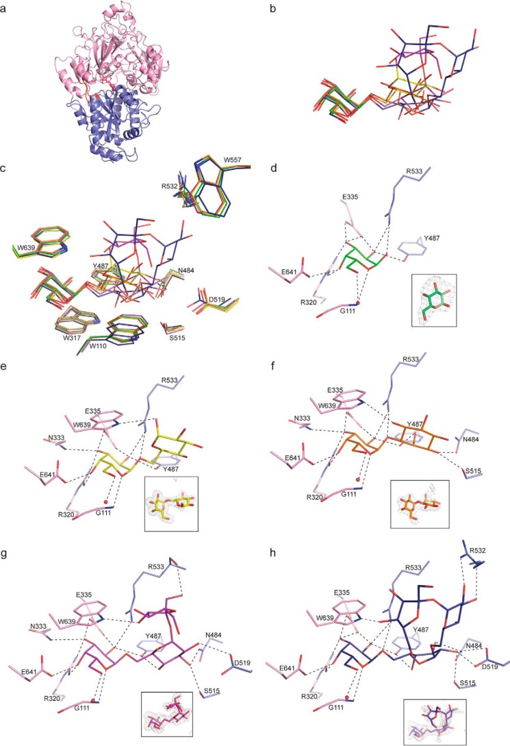
Figure 3.
Ribbon representation of MelB structures and ligand-binding site. a, raffinose in magenta is located in the cleft between lobes 1 and 2 shown in slate and in pink, respectively, and the hinge region is in red. b, superposition of the bound galactose, melibiose, galactinol, raffinose, and stachyose shown in green, yellow, orange, magenta, and blue sticks, respectively, in the binding site of MelB. c, same figure as in b showing the stacking between ligands and tryptophan (Trp639, Trp317, and Trp110). Except Trp639 and Trp317, all the other labeled amino acids mainly from lobe 2 can move up to 1 Å upon ligand binding. d–h, galactose (d), melibiose (e), galactinol (f), raffinose (g), and stachyose (h) bound to the binding site of MelB are shown in the same code color as in b. Hydrogen bonds between MelB and each ligand are shown as dashed lines in black (distances are up to 3.2 Å). A water molecule forming a hydrogen bond with each ligand is shown as a red circle. Each ligand is shown in its annealing Fo-Fc omit map contoured at 4σ.

