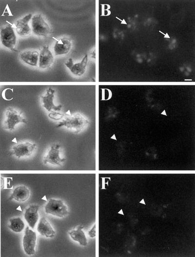
Figure 1.
PI 3-kinases regulate macropinocytosis. Wild-type cells (A and B), Δpik1/pik2 mutants (C and D), and cells treated with 20 μM LY294002 for 20 min (E and F) were bathed in growth medium containing RITC-dextran (2 mg/ml) for 5 min. A, C, and E are phase contrast, whereas B, D, and F are fluorescence images. The coverslips with the attached cells were washed and quickly fixed in 1% formaldehyde. Cells were viewed with the use of a fluorescence microscope, and images were captured with the use of T-MAX black and white 400 speed film. Exposure times were identical for all cells. Arrows in A and B denote cells with macropinosomes. Arrowheads in C–F denote cells without macropinosomes. Bar, 5 μm.

