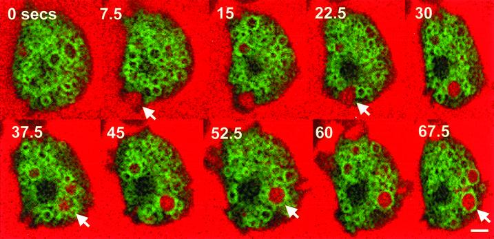
Figure 10.
GFP-Rab7 does not localize to forming macropinosomal cups. Cells expressing GFP-Rab7 were allowed to attach to coverslips and bathed in HL5 containing Texas Red dextran. Images were captured of a single cell undergoing macropinocytosis at 2.5-s intervals by confocal microscopy as described in MATERIALS AND METHODS. Arrows denote a large forming macropinosome that becomes ringed with GFP-Rab7. Bar, 5 μm.

