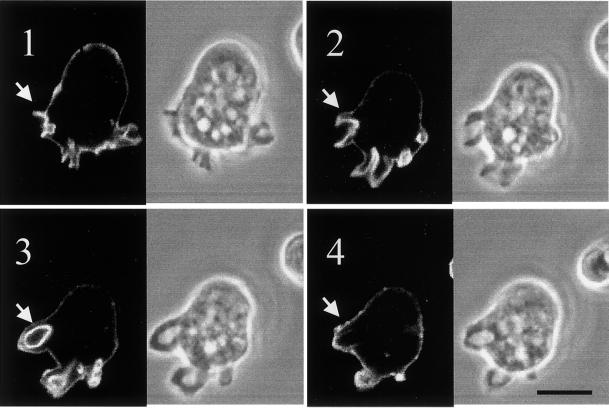
Figure 6.
GFP-ABD is recruited to macropinosomal cups. Cells expressing GFP-ABD and attached to coverslip images were captured by confocal microscopy as described in MATERIALS AND METHODS. Digital phase-contrast and fluorescence images were collected every 5 s. The montage shown in the figure represents images collected at 15 s (panel 1), 30 s (panel 2), 45 s (panel 3), and 60 s (panel 4). The arrow points to a forming and eventually complete macropinosome. Bar, 15 μm.

